

Welcome guest
Monday to Friday8AM - 9PM
AddressABC A-504, Rajnigandha Apartment, VADODARA-390007, GUJARAT, India.

Cardiac color duplex sonography is a medical imaging technique used to visualize the structure and function of the heart.
Cardiac color duplex sonography, also known as echocardiography or cardiac ultrasound. It employs high-frequency sound waves (ultrasound) to create real-time, dynamic images of the heart and its blood flow. The "color duplex" aspect of the technique refers to the use of color coding to display the direction and velocity of blood flow within the heart and its associated blood vessels.
The primary types of Cardiac color duplex sonography include:
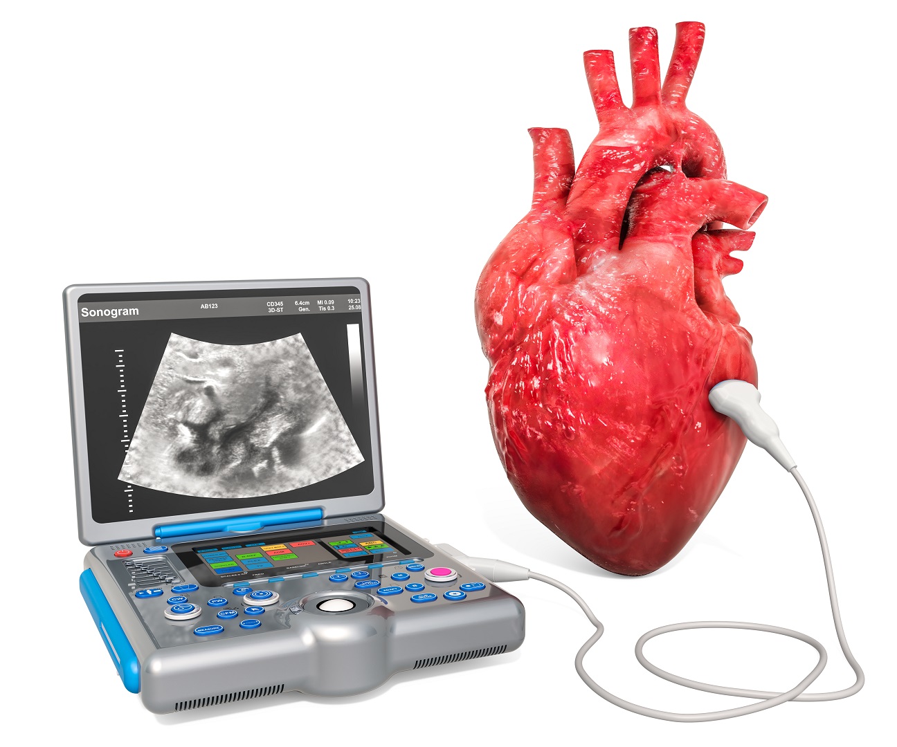
Following are the procedure steps:
Patient Preparation: The patient is typically asked to change into a hospital gown to allow easy access to the chest area. ECG electrodes are attached to the patient's chest to monitor heart rhythm during the procedure and lying on an examination table on their left side, to optimize imaging quality.
Application of Gel: A special ultrasound gel is applied to the chest area overlying the heart which helps transmit sound waves and improve the quality of the ultrasound images.
Placement of the Transducer: The healthcare provider places a transducer on the patient's chest. The transducer emits and receives high-frequency sound waves to create images of the heart.
Image Acquisition: The sonographer moves the transducer across the chest to obtain different views of the heart. These views include standard 2D images, as well as color Doppler images to visualize blood flow.
Doppler Imaging: Doppler ultrasound is used to assess blood flow patterns and velocities within the heart and blood vessels. The sonographer may use color Doppler to visualize blood flow direction and speed, and spectral Doppler to measure blood flow velocities at specific locations.
Image Recording: The images and Doppler data are recorded and stored for later analysis and interpretation by a cardiologist.
Completing the Examination: Once the necessary images and measurements are obtained, the sonographer may remove the ECG electrodes and wipe off the ultrasound gel from the patient's chest.
Interpretation and Reporting: The recorded images and data are reviewed and analyzed by a cardiologist or a qualified medical professional. A formal report is generated based on the findings, which is used for diagnosis and treatment planning.
The entire procedure typically takes about 30 to 60 minutes, depending on the complexity of the examination. It is a safe and non-invasive way to assess cardiac health and diagnose various heart conditions. Cardiac color duplex sonography plays a crucial role in cardiology by providing valuable insights into the structure and function of the heart.
India has become a popular destination for Cardiac Color Duplex Sonography due to its world-class medical facilities, experienced surgeons, and lower costs compared to other countries. Indian hospitals have state-of-the-art technology and equipment and offer excellent healthcare services at an affordable price. Patients from around the world can benefit from the cost-effective and high-quality healthcare services offered in India.
Gomedi9 is a medical tourism company that specializes in helping patients plan and organize their medical travel to India. They provide a range of services to make the process of medical tourism hassle-free and convenient for patients.
Gomedi9 can help patients to choose the right hospital, book accommodation, arrange transportation, provide interpreters, and ensure that the patient receives the best possible medical treatment at an affordable price. They have a team of experienced professionals who guide patients throughout the process of medical tourism, from the initial consultation to post-operative care.